




The Hospital de Clínicas of the University of Campinas (HC/Unicamp) celebrated, on April 10, a historic milestone for Brazilian public healthcare with the delivery of its new and fully restructured Nuclear Medicine Service (SMN) area.
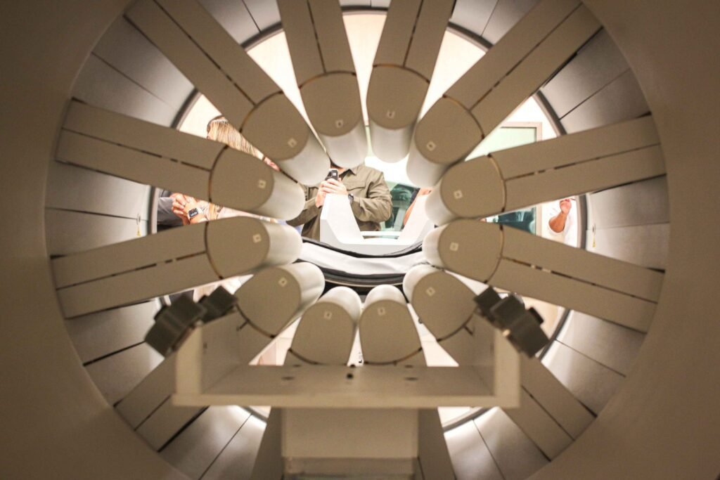
One of the major highlights of the renovation was the acquisition of a SPECT/CT (Single Photon Emission Computed Tomography combined with Computed Tomography) system featuring CZT (Cadmium, Zinc, and Tellurium) detector technology, making HC/Unicamp the first public hospital in Latin America to have this technological innovation. This acquisition allows patients of the Brazilian Unified Health System (SUS) to access high-precision three-dimensional imaging exams performed up to four times faster.

Investments in the modernization of the SMN
The completion of the restructuring and the acquisition of the SPECT/CT CZT equipment relied on decisive support from the Legislative Branch. The securing of approximately R$ 4.5 million in parliamentary amendments was essential for this, a joint effort supported by federal deputies Paulo Freire, Kim Kataguiri, and Adriana Ventura, as well as former federal deputy Cátia Sastre.
The approximate cost of the equipment, transportation, and installation exceeded R$ 8.6 million, partially funded by these parliamentary amendments and the remaining portion by the São Paulo Research Foundation (FAPESP), through the project that enables CancerThera—supported by the foundation as one of its Research, Innovation, and Dissemination Centers (CEPIDs)—also counting on funding from the Unicamp Rectory.
In an interview, Dr. Carmino Antônio de Souza, an onco-hematologist, professor at the School of Medical Sciences at Unicamp, principal investigator at CEPID CancerThera, and vice president of FAPESP, revisited the trajectory of restructuring the hospital’s imaging area, which began in 2012 but reached a new level of excellence with the start of the research center’s activities.
According to Souza, the initial milestone of this major transformation in the imaging area was the acquisition of a PET/CT (Positron Emission Tomography combined with Computed Tomography) system, also funded at the time by FAPESP through a Multi-User Equipment (EMU) call. This first achievement represented a fundamental leap in quality for the institution, boosting thesis production, academic research, and oncological care, and highlighted the ongoing need to improve the hospital’s diagnostic infrastructure.
To maintain excellence and integrate the complex with new demands, also with FAPESP resources via CancerThera, a major technological upgrade of the same equipment was secured, requiring an investment of approximately R$ 2.3 million. With this renewal, the system now offers tools that enable, for example, dynamic PET/CT exams and PET-guided biopsies, among other features that dramatically increase diagnostic precision.
“The restructuring of Nuclear Medicine is a fundamental element for us to fully carry out the project, especially because at every stage of the theranostic approach—whether in diagnosis, in defining the carrier, tumor avidity, or antitumor action—we fundamentally depend on having high-quality imaging at all these stages,” the researcher explains.
Souza also emphasized the inherent mission of the research center he coordinates to serve the state of São Paulo and the country: “Our work at CancerThera is linked to major teaching and research institutions, which are public entities working exclusively for patients in the public health system.” He concluded by highlighting the university’s broader goal: “HC/Unicamp must be at the forefront. We must seek every possible means to fulfill this role in Medicine and Health Sciences at the frontier of knowledge.”


Researchers from CancerThera celebrate the acquisition of the SPECT/CT CZT and the restructuring of the SMN/HC/Unicamp (including the construction of a therapeutic room), which will benefit the center’s clinical research in the theranostic model in cancer. In the photo, from left to right: M. Elvira Corrêa, Carmino A. de Souza, Fabio L. N. Marques, Carmen S. P. Lima, Victor M. Deflon, Luciana M. Quaglio, Elba Etchebehere, Pedro P. Corbi, Bárbara J. Amorim, and Celso Dario Ramos.
Inauguration ceremony with the presence of supporters
The new space combines personalized medical care, advanced scientific research in theranostics, and innovation in cancer treatment. The transformation was detailed by the coordinator of the SMN/HC/Unicamp, Dr. Bárbara Juarez Amorim, who described past operational challenges, including inadequate infrastructure and equipment operating at the end of its lifespan. In her presentation during the inauguration ceremony, Amorim explained that collective efforts—combining parliamentary funding, CEPID CancerThera technical reserve, and support from the Unicamp Rectory—changed this scenario.
The new physical restructuring, valued at R$ 2.1 million, encompassed much more than the main equipment room. “We installed a therapeutic room, which is essential for performing theranostic procedures, and also upgraded our radiopharmacy—the heart of Nuclear Medicine,” highlighted the nuclear physician, who is also an associate researcher at CancerThera.
The convergence between scientific funding and direct impact on citizens’ lives in the state of São Paulo was emphasized by the president of FAPESP, Dr. Marco Antônio Zago, a clinician and professor at the Ribeirão Preto Medical School of the University of São Paulo. “This project is an example of how FAPESP uses the funds it receives,” he stated. “The population also wants to see this money applied to science and technology projects that have an immediate effect on improving quality of life.”
For Zago, the magnitude of CancerThera lies in its ability to bridge theoretical development and SUS patient care. “Centers [like CancerThera] operate between basic science and applied Brazilian science, making this transition, with people working on the most fundamental aspects of cancer biology while also inaugurating an imaging machine that will serve both research and patient care,” he added.
The enhancement in healthcare delivery brought by this modernization was highlighted by Unicamp’s general coordinator, Dr. Fernando Antônio Santos Coelho, an industrial pharmacist and professor at the Institute of Chemistry. According to him, the SPECT/CT CZT will make a difference “not only for research development but especially for more accurate and earlier cancer diagnoses.” He added: “Once a public hospital has high-complexity technology, it has the commitment to serve a large portion of the population, continuously improving the quality of what it delivers.”
At the closing of the ceremony, Dr. Paulo César Montanher, physical educator, professor at the School of Physical Education, and rector of Unicamp, praised the academic team’s efforts and recalled the historical character of the area. “Our Nuclear Medicine Service has a pioneering tradition of more than 30 years, with a continuous search for solutions and advancements,” he noted. He also emphasized the long-term legacy beyond care: specialized education for university students.





LEARN MORE | Technological advantages of the new SPECT/CT CZT equipment
The SPECT/CT exam is a high-technology Nuclear Medicine imaging procedure, mainly used for diagnosing and monitoring cancer patients. SPECT stands for Single Photon Emission Computed Tomography and CT refers to Computed Tomography.
The equipment works by fusing scintigraphy with tomography, generating full-body three-dimensional (3D) images. This combination integrates functional and anatomical imaging, allowing physicians to detect tumor and metastasis locations with extremely high accuracy.
The newly inaugurated model features CZT technology (Cadmium, Zinc, and Tellurium), using detectors arranged 360 degrees around the patient. Combined with Artificial Intelligence, this makes the exam up to four times faster than conventional models, offering excellent resolution and greater comfort.
This combination of speed and high image quality is essential for precision medicine, particularly to support theranostics (an approach that combines diagnosis and therapy in a single process), ensuring more accurate diagnoses, targeted therapies to destroy tumor cells, and greater patient comfort during procedures.
Text and photos: Romulo Santana Osthues