




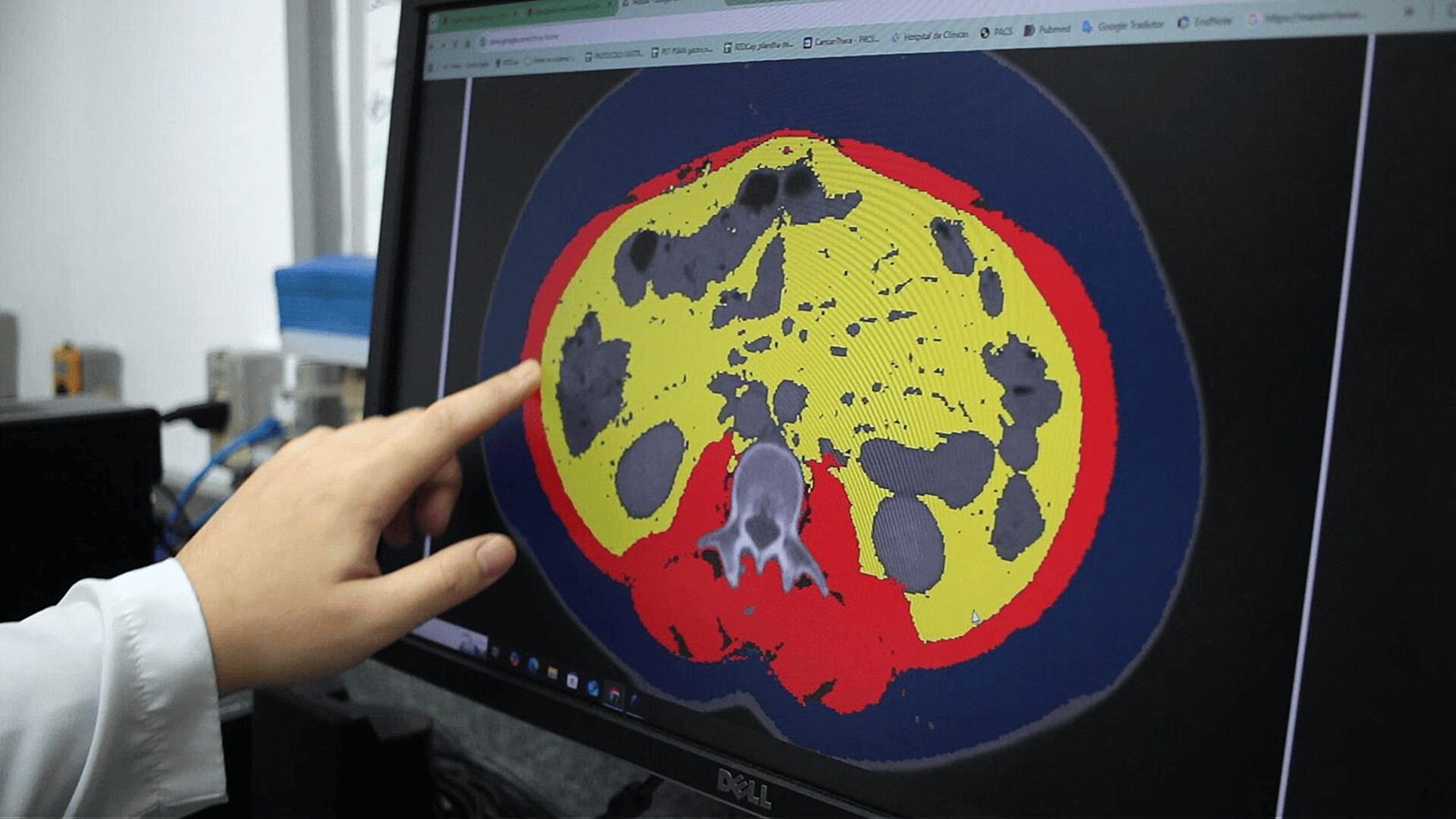

A multidisciplinary team, combining expertise from Physics, Oncology, and Nutrition, developed an unprecedented mathematical model based on Artificial Intelligence (AI) in the laboratories of the University of Campinas (Unicamp). The result is a computer program capable of mapping, in detail, the body composition of oncology patients in less than one minute, potentially supporting clinical practice and precision medicine by providing more accurate prognoses and personalized therapeutic guidance.
The idea arose from the need to optimize the interpretation of computed tomography (CT) scans, which are essential tools in monitoring cancer patients. Until the development of this program, mapping body tissues in these radiological images was a time-consuming and entirely manual task for researchers.
Dr. Jun Takahashi, a nuclear physicist, full professor at the Gleb Wataghin Institute of Physics (IFGW) at Unicamp and an associate researcher at CEPID CancerThera, reports that manual evaluation performed by the medical team collaborating on the project took between 30 and 60 minutes to complete the analysis of each image. In addition to being slow, the traditional method was subject to significant variability. Accuracy depended, for example, on the professional’s level of fatigue, time of day, or degree of attention during image interpretation.
“If we considered the time researchers take to perform manual analyses, it would take two years to deliver the data,” recalls the physicist, referring to the moment when a partner Oncology team requested the evaluation of a large image database containing approximately 500 scans. Faced with this scientific and clinical bottleneck, the researchers decided to develop an automated solution.
The software is not yet available to the general public and depends on licensing interest from healthcare companies to reach the market. Currently, it is used only in a research environment.
How the program works
The program operates through a process known as “image segmentation.” When a patient undergoes a CT scan, the resulting image displays bones, organs, muscles, and fat, all mixed within a complex grayscale scale. The AI-based software developed by Takahashi and his team scans this image and creates a digital mask, autonomously and precisely separating and color-coding each tissue type.
What distinguishes this code from other generic models available online is its rare ability to accurately identify and separate muscle tissue and three specific types of fat: subcutaneous adipose tissue (fat just beneath the skin), visceral adipose tissue (fat accumulated between organs), and intramuscular adipose tissue (fat interspersed within muscle fibers). The analysis is based on CT images at the level of the third lumbar vertebra (L3).
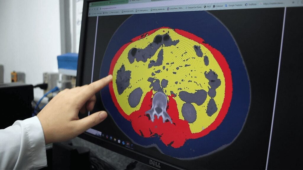
known as L3.
“On the internet, you will find many codes that use CT images to perform segmentation, identifying, for example, the liver or kidneys. But separating these three adipose tissues—intramuscular, subcutaneous, and visceral—you won’t find,” he emphasizes. The program also identifies and isolates bone structures to avoid interference in the results.
The algorithm’s high accuracy—with an estimated error margin of less than 5%, even in difficult tissues such as intramuscular fat—is not due solely to computational power. The key differentiator was the integration of more than a decade of clinical experience from the collaborating medical team directly into the mathematical architecture of the code.
During AI training, researchers observed that the algorithm initially confused bone marrow with muscle tissue due to their similar grayscale intensity measured in Hounsfield Units (HU). By incorporating human reasoning logic from nutritionists and physicians, the system learned to exclude bones before searching for fat tissues. “We embedded this experience into the code architecture. It differs from typical machine learning approaches that simply feed data into the system,” says Takahashi.
Another key finding involved radiological calibration. Researchers discovered that precise calibration of grayscale intensity reveals clinically relevant information about tissues. Incorporating this code directly into CT scanners could represent an innovation for the imaging equipment industry, enabling patients to receive automatic body composition reports immediately after the scan.

Initially, the input image undergoes a preprocessing stage with the application of a Hounsfield Unit (HU) filter. Next, U-Net–based models combined with ResNet18 perform tissue segmentation, classifying muscle, intramuscular adipose tissue, visceral adipose tissue, and subcutaneous adipose tissue. Finally, a new HU filter is applied in post-processing, generating the final color-coded segmented image. The diagram was created by the researchers.
Interdisciplinary and multidisciplinary collaboration
This synergy between human knowledge and program operation ensures process uniformity, which is crucial for scientific reliability. Understanding uncertainty in medical exams is vital, as quantitative variation can significantly impact treatment dosage or therapeutic decisions.
In cancer treatment, this AI-based technology opens promising new avenues. Dr. José Barreto Campello Carvalheira, an oncologist, full professor at the School of Medical Sciences (FCM) at Unicamp and principal investigator at CancerThera, leads studies on cancer immunometabolism, a field investigating the systemic interaction between the disease and the patient’s body.
He explains that oncology staging traditionally focuses almost exclusively on tumor characteristics. However, the patient’s metabolic response to the tumor is equally determinant for survival.
“One of the main limitations in oncology is the difficulty in accurately estimating patient prognosis based solely on tumor characteristics. Body composition studies expand this understanding by enabling assessment not only of tumor–host interaction but also of the patient’s capacity to respond to disease and treatment,” he explains.
With the program analyzing over 500 patient images in just 30 minutes, specialists can rapidly observe how different tumor characteristics affect the body. This computational speed allows medicine to begin identifying new patient subtypes, potentially sparing individuals from aggressive or ineffective treatments.
“Does the patient really need chemotherapy? When we identify that a tumor belongs to a less aggressive group, we can consider less intensive therapeutic strategies,” the oncologist projects.
The program’s impact also extends strongly to Clinical Nutrition. Dr. Maria Carolina dos Santos Mendes, a nutritionist and postdoctoral researcher in Research Management—Innovation and Technology Transfer associated with CancerThera, highlights the limitations of current hospital tools, which rely heavily on simple measurements such as waist and calf circumference.
“In clinical practice today, we simply cannot obtain this level of information. It would greatly help if CT reports included body composition analysis alongside tumor data,” she notes. Access to mathematically precise data on fat and lean mass will enable highly targeted nutritional interventions and early clinical action.
Software registration and market implementation
The technology’s future is promising, and next steps are already defined. The software has been formally registered with strategic support from Inova Unicamp. Prior to registration, a preliminary market analysis assessed its application potential.
Now protected, the agency is actively seeking partners and presenting the technology to industry, evaluating the need for commercial adaptation or technological maturation.
According to Ma. Beatriz Hargrave, technology transfer supervisor at Inova Unicamp, “partnerships can be established for co-development, aligning the technology with partner needs and increasing the chances of large-scale adoption.”
There is strong expectation of industry interest, as the program addresses a real and urgent healthcare demand, with potential applications in hospitals, clinics, and imaging centers due to efficiency gains, reduced analysis time, and improved result consistency.
The success and precision of the technology stem directly from academic collaboration. Integrating Physics, Oncology, and Nutrition enables robust innovations aligned with real-world clinical needs.
At this stage, research teams from IFGW/Unicamp and FCM/Unicamp are developing a user-friendly interface: a clean screen with interactive buttons allowing healthcare professionals to visualize muscle or fat layers with a single click.
NOTE FOR INTERESTED PARTIES: The transfer of protected technologies from Unicamp, such as this software (“PC346_SEGMENTATION”), and the formalization of R&D&I agreements are supported by Inova Unicamp. Interested organizations should contact the agency through its official form.
Text and photo: Romulo Santana Osthues