




In recent years, the way Oncology classifies the severity of a cancer has gained a new resource thanks to the development of computer programs with Artificial Intelligence (AI) applied to the medical field.
A recent study, for example, conducted by researchers at the University of Campinas (Unicamp), demonstrated that the automated analysis of body composition of patients with gastric (or stomach) cancer is capable of accurately predicting mortality risks when specific characteristics of muscles and fats are measured through Computed Tomography (CT) scans.
The study was published in the scientific journal Informatics in Medicine Unlocked (volume 52, 2025) under the title Improving resectable gastric cancer prognosis prediction: A machine learning analysis combining clinical features and body composition radiomics, being conducted by researchers from the Gleb Wataghin Institute of Physics (IFGW) and the School of Medical Sciences (FCM) at Unicamp – including those from CEPID CancerThera.
What the standard staging system does not see
Staging (or performing staging) is the medical process of assessing the severity and extent of a cancer in the patient’s body in order to try to predict how the disease will evolve and help healthcare professionals define the best treatment.
Historically, Oncology relies on a staging system known as TNM to predict clinical outcomes. The acronym refers to Tumor (size and extent of the main tumor), Node (indicating whether cancer has reached the lymph nodes), and Metastasis (indicating whether the disease has spread to other organs).
Because it does not evaluate body composition, TNM alone cannot capture the full diversity of ways cancer manifests, which may result in patients classified at the same stage of the disease (such as Stages II or III) presenting very different disease progressions and survival times. This lack of precision can lead to situations in which severity is underestimated by the TNM system or, conversely, may cause overtreatment in patients who would be low risk.
“What we have shown in our line of research and in this study in more detail with AI is that TNM is only part of the story,” explains Dr. José Barreto Campello Carvalheira, oncologist, full professor at FCM/Unicamp and principal investigator at CancerThera. “By incorporating body composition – especially quantitative characteristics of muscle and fat extracted by radiomics – we begin to stage not only the tumor, but the patient as a whole.”
To reach these conclusions, the team used data and tomographic images (at the level of the third lumbar vertebra – L3) from 276 patients with gastric cancer treated at the Unicamp Clinical Hospital between 2009 and 2018. Through machine learning algorithms, the computer program developed with AI analyzed the images and extracted complex radiomic data on body composition (especially muscles and fats).
What is radiomics?
It is a technique that uses data characterization algorithms to extract, quantify, and analyze detailed information from medical imaging exams, such as CT scans. It acts as a non-invasive biomarker, allowing computers to comprehensively quantify the physical and structural characteristics of a tumor or a patient’s body.
Radiomics evaluates the distribution of pixel values and can compute numerous mathematical and statistical characteristics. Among the metrics extracted by this method are: percentiles of the grayscale or radiodensity scale (such as the 10th and 90th percentiles mentioned earlier); entropy, energy, and variance; and median, skewness, and kurtosis.
The algorithm cross-referenced body composition with clinical data and identified that patients in Stage II (theoretically less severe), but who had a body classified by AI as “high risk,” had a life expectancy identical to that of patients in Stage III. “This is potentially disruptive because it explains a classic limitation of Oncology: patients at the same stage have very different outcomes,” notes the physician. “In other words, there is relevant biological heterogeneity that TNM alone does not capture,” he adds.
Body mass and disease progression
When body structures are analyzed with precision (through tomography and radiomics), indirect signs of important processes can be identified. The study revealed that the extremes of density (the 10th and 90th percentiles) of muscle and visceral fat were the strongest predictors of survival. Patients with abnormally high radiodensity in visceral fat showed greater weight loss, lower Body Mass Index, and consequently saw their median survival time drop by half.
The changes observed by healthcare professionals function as a snapshot of serious processes, such as chronic inflammation (which may favor tumor growth) and cachexia (a syndrome of extreme weight and muscle loss that debilitates the patient and reduces their ability to tolerate chemotherapy). “Although the cancer is in the stomach, it is not an isolated disease – it interacts with the entire organism. Muscles and fats are metabolically active tissues and reflect the body’s ‘internal state’,” details José Barreto.
The researcher summarizes the biological dynamics of the disease: “The tumor is the problem, but the state of the body defines how much the patient can respond to it. AI simply helps us measure this with much greater precision than was previously possible.”
A leap in scale and the role of AI
Until recently, performing this tissue segmentation analysis image by image was an exhaustive, time-consuming, and strictly manual task. With the new AI program, the operational bottleneck no longer exists. “In fact, the algorithm allows body composition analysis to be carried out on a large scale,” highlights physicist Gianni Shigeru Setoue Liveraro, PhD candidate at IFGW/Unicamp and lead author of the study published in Informatics in Medicine Unlocked.
“In the medical context, AI has been used mainly as a tool: it performs specific and well-defined tasks but does not make decisions about what should be done. Decisions continue to be made by qualified professionals,” clarifies the researcher. In Liveraro’s view, the goal of the developed program is to optimize clinical routine: “The results should be more accurate diagnoses and more effective treatments, leading to a better quality of life for patients.”
Despite the promising results, the scientists involved in the development of the computer program used for segmentation and its medical application remain methodologically cautious. The program, which has already been registered with the support of the Innovation Agency Inova Unicamp, will still need to be tested in other prospective clinical studies and across multiple healthcare centers.
Currently, the data provided by the program should only be used to classify the risk level of each patient with stomach cancer, helping to predict who is more likely to experience complications. At this stage, the technology does not serve to determine direct changes in the main oncological treatment – such as altering chemotherapy doses or indicating different surgeries.
Gastric cancer, according to the National Cancer Institute (Inca), is the third leading cause of death from malignant tumors worldwide, and even after modern treatments, it shows high recurrence rates.
In daily hospital practice, considering that the recurrence rate in gastric cancer is high and early detection remains a challenge, the program can function as an important warning signal, indicating which patients are physically more fragile and therefore require rigorous and frequent monitoring. Based on this alert, the healthcare team can act preventively in areas that are fundamental for survival but are often secondary in traditional treatment.
This includes offering immediate nutritional support, attempting to curb excessive muscle mass loss, and controlling systemic inflammation in patients identified as more vulnerable. Only through future studies will healthcare teams be able to determine whether adjusting medications based on these data will help increase patients’ survival time.
“We still do not know whether changing management based on this type of model improves survival. This is precisely the question that needs to be answered in prospective studies. There is even a bidirectional scenario to be tested – both intensifying treatment in patients ‘underestimated’ by TNM and avoiding overtreatment in low-risk patients,” explains oncologist José Barreto.
Inside the program: how AI sees the patient’s body
The major strength of the computer program used is a mathematical model based on Artificial Intelligence (AI) focused on automated reading of Computed Tomography (CT) scans. The main innovation of the tool lies in its level of detail when analyzing muscle and adipose tissues.
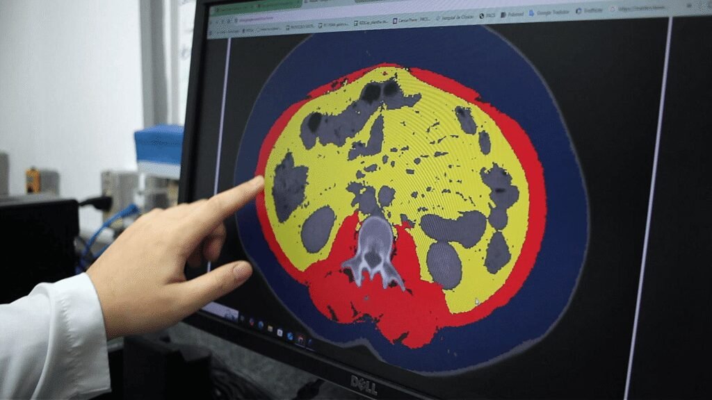
While generic algorithms usually identify larger organs, the program developed at Unicamp (with U-Net and ResNet18 models) is able to map, from the CT image at the level of the third lumbar vertebra (L3), and meticulously isolate the patient’s muscles and fat into three distinct categories: subcutaneous (located just below the skin), visceral (accumulated between the organs), and intramuscular (interspersed within muscle fibers). The program creates a mask on the image, separating these tissue types and coloring each one differently – the original images are generated in grayscale by the CT scanner.
The program’s success was achieved by incorporating hospital experience directly into the programming. The AI learned to read the images by adopting the clinical reasoning of physicians and nutritionists. “We brought 10 years of the medical team’s experience into the code architecture,” explains Dr. Jun Takahashi, nuclear physicist, full professor at IFGW/Unicamp, associated researcher at CancerThera, and responsible for the team that developed the program.
The technology solved a major bottleneck in radiomics research: time. Tissue mapping, which previously required 30 minutes to one hour of meticulous manual work by the medical team, is now completed “in less than a minute,” highlights the physicist. In addition to speed, which eliminates variations caused by natural human fatigue and enables research with large data volumes, there is a gain in precision and uniformity in the process, which is very important for scientific practices. With the ability to generate instant reports of body composition, the tool paves the way for highly personalized interventions.
At this link, you can learn the details of the development and operation of the program.
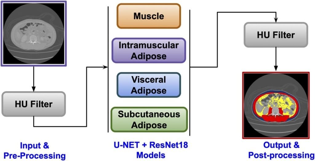
Initially, the input image undergoes a preprocessing step with the application of a Hounsfield unit (HU) filter. Then, models based on U-Net combined with ResNet18 perform tissue segmentation, classifying muscle, intramuscular adipose tissue, visceral adipose tissue, and subcutaneous adipose tissue. Finally, a new HU filter is applied in post-processing, generating the final segmented colored image. The diagram is authored by the researchers.
Text e photo: Romulo Santana Osthues