
Positron emission tomography (PET) and computed tomography (CT) are imaging exams widely used in healthcare for detailed and non-invasive internal body analysis.
PET allows the evaluation of both the anatomy and metabolism of tissues and organs, identifying possible abnormalities. CT highlights the body’s structure using X-rays and computer processing, generating high-resolution images in multiple planes.
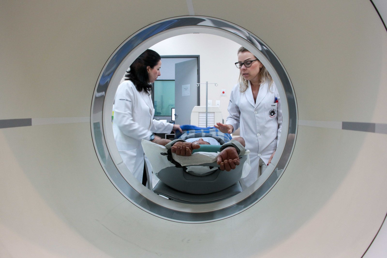
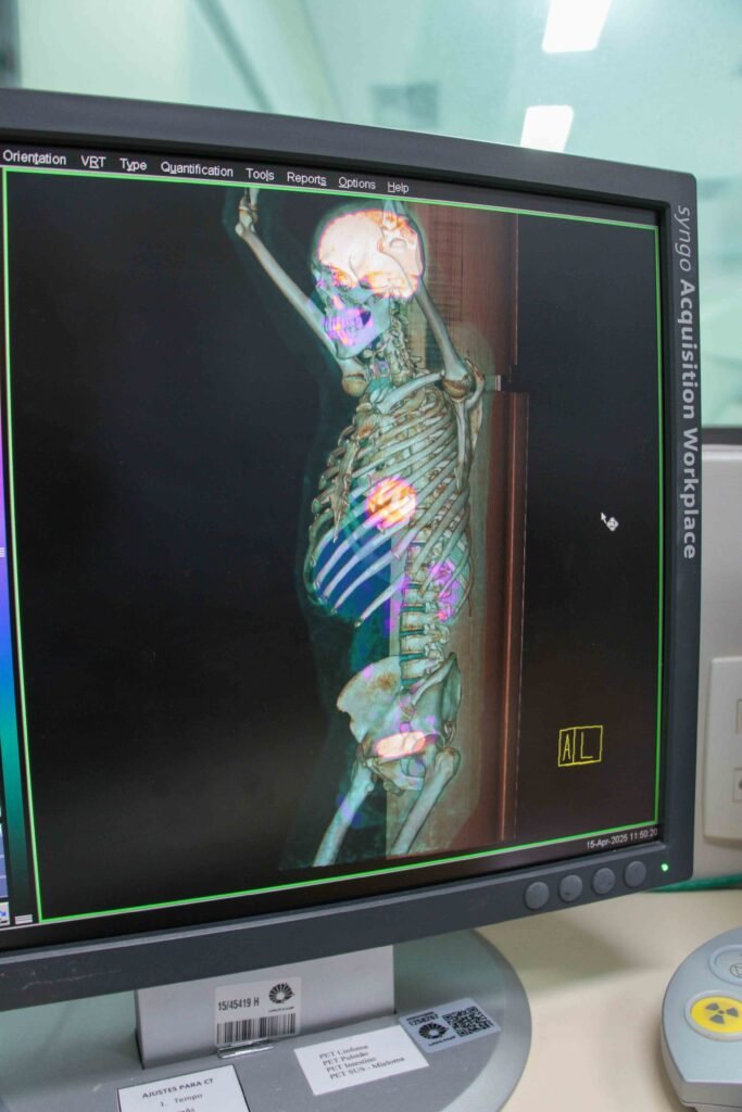
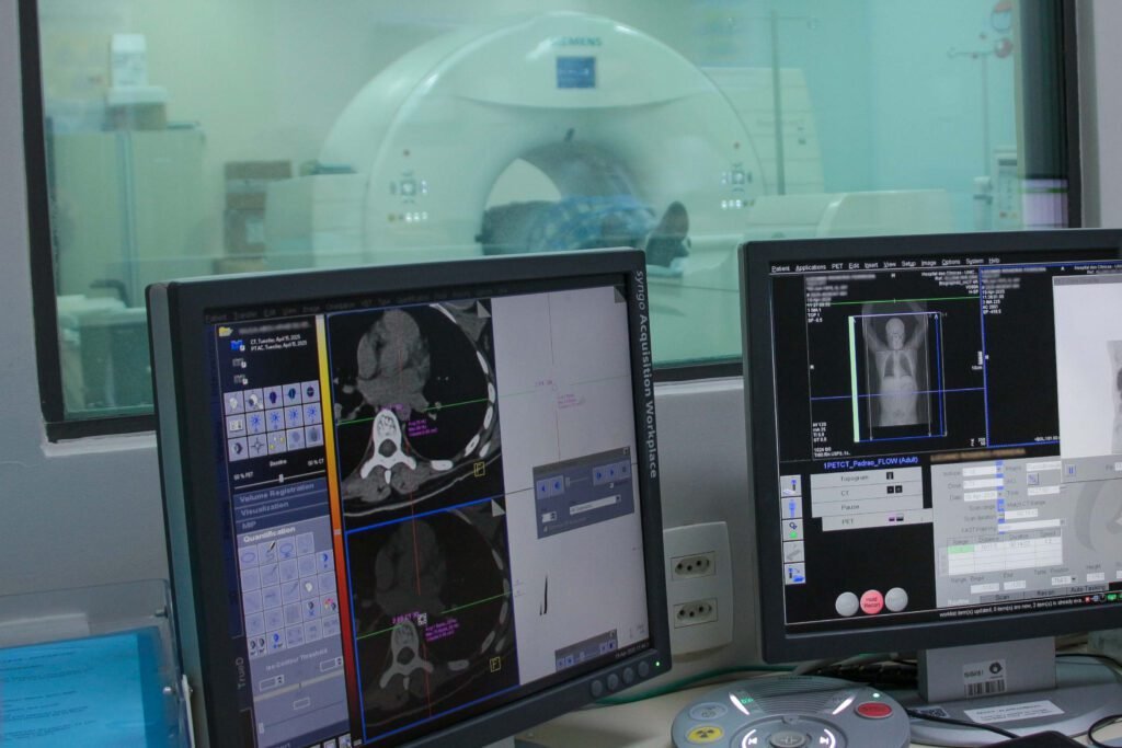
These exams can be combined in a single device known as PET/CT. After the administration of radiopharmaceuticals to the patient, the equipment produces 3D images that allow investigation of the body’s structure and function as a whole.
One such device is used by the specialists and researchers at the Cancer Theranostics Innovation Center (CancerThera), a Research, Innovation, and Dissemination Center (CEPID) based at the University of Campinas (Unicamp). With funding from FAPESP, a recent upgrade of the PET/CT system was made possible, enhancing its systems and adding new features and accessories.
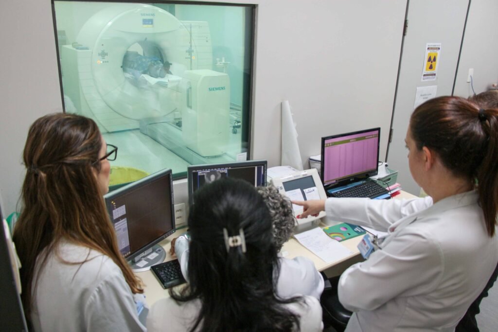
Installed in 2013 at the Nuclear Medicine Service of Unicamp’s Clinical Hospital (SMN-HC), the Biograph mCT PET/CT scanner by Siemens is used by nuclear medicine and oncology teams for detecting tumors and metastases, as well as by CEPID researchers conducting preclinical and clinical studies.
In addition to testing new uses for well-established metallopharmaceuticals and radiopharmaceuticals in specific cancers, CEPID CancerThera researchers are working to develop new “theranostic” applications—a concept that integrates therapy and diagnosis in a single approach.
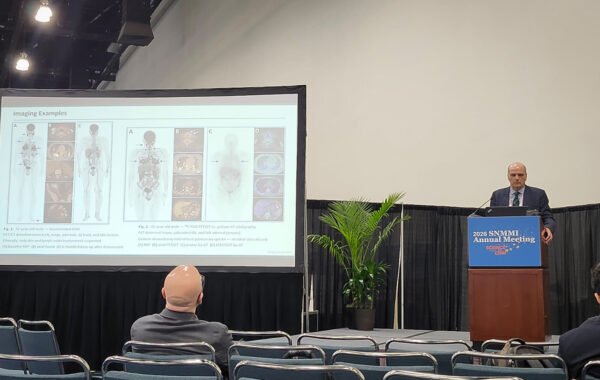
“PET/CT has become one of the most important exams in medicine and science, not only for cancer but also for many other diseases, including inflammatory, infectious, autoimmune, and metabolic conditions,” says Carmino Antonio de Souza, hemato-oncologist, professor at Unicamp’s School of Medical Sciences (FCM), lead researcher at CEPID CancerThera, and vice president of FAPESP.
“PET/CT is a key element in theranostic research. New radiopharmaceuticals developed at CEPID CancerThera, labeled with positron emitters, will continue to be evaluated using this equipment—now, with improved image quality,” says Celso Dario Ramos, nuclear medicine physician, professor at FCM-Unicamp, and lead researcher at CEPID CancerThera.
“We’re not the only nuclear medicine center in the country with these features, but as a PET-focused training and research facility, I’d say we’re one of the best equipped,” Ramos adds.

Key new features
In April this year, the PET/CT system was enhanced with faster and more efficient processing and quantification tools based on artificial intelligence (AI), which also enabled the increase in software licenses from three to six—allowing more users to simultaneously generate and analyze images.
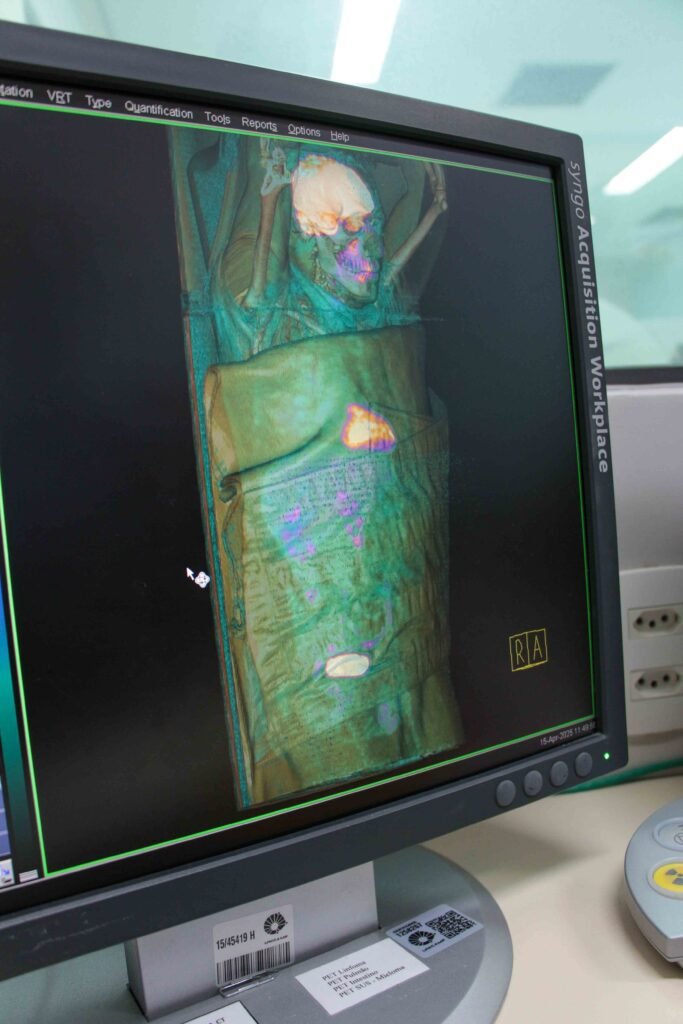
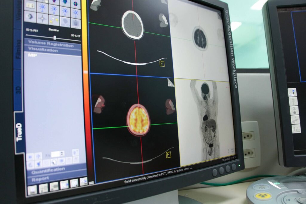
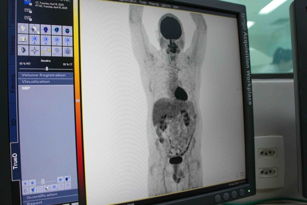
Previously, PET images were acquired in segments; now, image acquisition can be continuous, simplifying exam planning and improving visualization quality. The system can also generate dynamic images over a pre-established time frame, allowing researchers to track how fast the radiopharmaceutical is absorbed by tissues.
In Nuclear Medicine, the term “uptake” refers to how intensively tissues absorb radiopharmaceuticals from the bloodstream. For example, in cancer, uptake may be high or low, and its distribution throughout the body can be visualized through PET/CT images.
“Using AI, even when image uptake points are minimal due to a lower radiation dose, the computer enhances the image quality, making it easier to detect lesions,” says Ramos.
Another AI feature enables the computer to automatically identify, delineate, and quantify normal and abnormal areas.
The upgrade also includes accessories for guided biopsies, during which a needle is inserted precisely where the PET/CT image shows a radiopharmaceutical uptake abnormality—allowing a clearer understanding of what’s occurring in that specific area. The procedure can be monitored in real time using new in-room screens.
Nuclear medicine physician Bárbara Juarez Amorim, coordinator of the SMN-HC-Unicamp and associate researcher at CEPID CancerThera, highlights guided biopsy as one of the biggest advantages of the upgrade, as it helps diagnose suspicious or unclear lesions. “Sometimes, PET alone cannot distinguish between cancer and something benign, like inflammation,” she explains.
Open access and funding
The PET/CT system at SMN-HC-Unicamp is part of FAPESP’s Multiuser Equipment Program (EMU) and is accessible to the entire scientific community of São Paulo state—appointments can be made in advance via phone (+55 19 3521-7772) or email (soniama@hc.unicamp.br).
Researchers will have access to PET/CT imaging using various radiopharmaceuticals, with enhanced visualization, processing, and guided biopsy features for both oncological and non-oncological research.
“FAPESP has worked hard to ensure that high-value equipment funded by the foundation is not restricted to a single researcher or group but can be used by a wide scientific community,” Souza emphasizes.
The total cost of the PET/CT upgrade was approximately USD 500,000, fully funded by FAPESP, which had also financed the equipment’s initial acquisition in 2013 (USD 2.4 million).
Choosing to upgrade rather than replace the equipment resulted in major cost savings. “A new PET/CT system with these features could cost up to USD 10 million,” says Souza. The upgraded equipment is expected to remain in good working condition for another ten years.
Text and photos: Romulo Santana Osthues | Originally published on Agência FAPESP portal.