




Paracoccidioidomycosis (PCM), neglected systemic mycosis of high clinical and epidemiological relevance in Brazil, gained a powerful ally in the precise mapping of its severity. A new study conducted by researchers from the CEPID CancerThera demonstrated that molecular imaging technology by FDG PET/CT is able to detect more than twice as many bodily injuries as compared to conventional staging methods.
The work, entitled FDG-PET/CT Demonstrations superior detection of multiorgan involvement in Paracoccidioidomycosis compared with conventional staging and published in the scientific journal Clinical Infectious Diseases, points out that the tool redefines the panorama of the disease, with direct impacts on the medical management and treatment time of patients.
Expanded accuracy against a challenging mycosis
Popularly known as the “coffee planter disease”, paracoccidioidomycosis is contracted by inhalation of spores of the genus fungus paracoccidioides present on the ground. Although it primarily affects the lungs, the infection can spread silently and aggressively to multiple organs via lymphatic and blood pathways. The great clinical challenge lies precisely in the traditional staging[processo médico que consiste em avaliar a extensão, a gravidade e o grau de disseminação de uma doença pelo corpo para definir a melhor conduta terapêutica]: physical examination, serology and chest X-rays often underestimate the true extent of the parasitic and inflammatory load in the body, identifying isolated lung lesions while extrapulmonary foci go unnoticed.
To solve this gap, the research prospectively evaluated 34 patients with a confirmed diagnosis of PCM (among untreated individuals and individuals with suspected therapeutic failure). All participants underwent both standardized clinical-conventional staging and full-body examinations with FDG PET/CT.
The results obtained impress: “In our study, we observed that the PET/CT identified more than twice as many lesions compared to the conventional evaluation (150 versus 62), detecting, for example, new areas of disease involvement that had not been diagnosed with other Examinations, with emphasis on lymph nodes, lungs and adrenal glands. In addition, the PET/CT reclassified patients as having multifocal disease in 97% of cases and increased the proportion of cases classified as severe, from 70.6% to 91.2%, showing that the method can be more accurate in evaluating both the extent and severity of the disease”, details the researcher Pricila Gama da Cunha, PhD student in Internal Medicine at the Faculty of Medical Sciences at UNICAMP (FCM-UNICAMP).
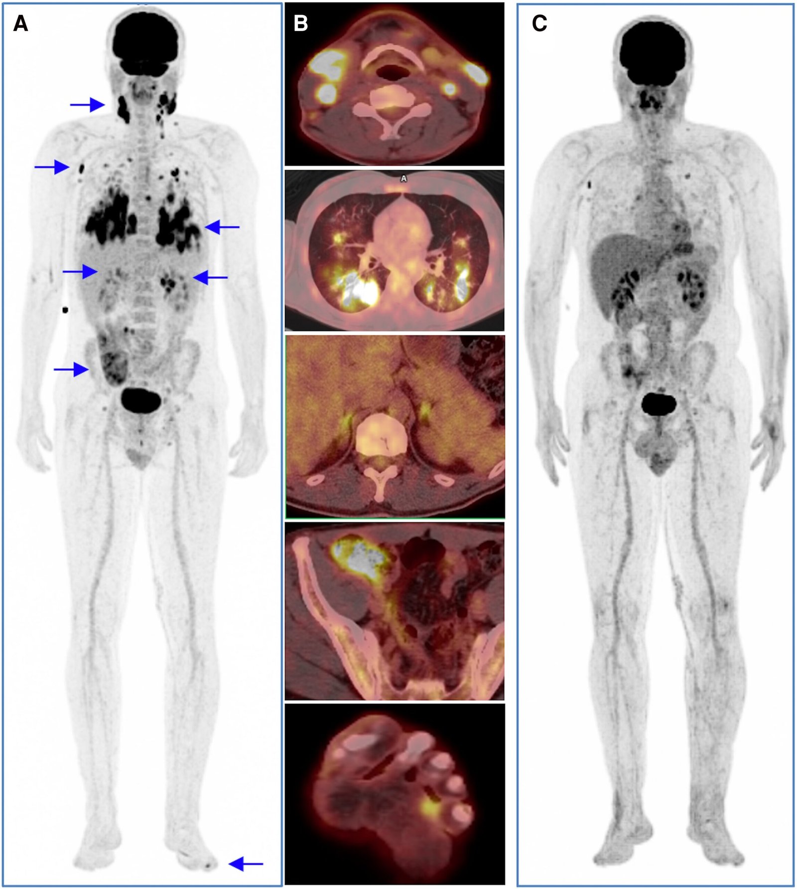
The reclassification of paracoccidioidomycosis as a multifocal and severe condition brings direct reflexes to medical practice. As explains Dr. Plinio Trabasso, an infectologist and associate professor at FCM/UNICAMP, this conceptual change improves the understanding of the pathophysiology of the infection[mecanismos biológicos e celulares pelos quais os agentes agressores provocam o desenvolvimento e a evolução de uma doença no organismo]. As the specialist points out, the systemic disease has a “very intense inflammatory component, which explains the consumptive syndrome that accompanies the condition and precedes the diagnosis” – a serious clinical picture, characterized by marked weight loss, malnutrition, extreme weakness and progressive physical debilitation.
What the exam sees and the value of the metabolic biomarker
To understand how doctors can assess the severity of paracoccidioidomycosis, it is necessary to look inside the body through technology. The PET/CT scan uses a radioactive marker called FDG — a molecule analogous to glucose that “illuminates” the sites in the body where there is an accelerated consumption of energy. As both cancer cells and the foci of serious infections and inflammation expend a lot of energy, they stand out in the exam.
This is where quantitative and volumetric biomarkers come in: such as the metabolic volume of lesions (TMLV) and total glycolysis (TLG), advanced metrics evaluated in the study. In simple terms, instead of just taking a static ‘photo’ to see where the infection is, researchers can calculate, through software, the total volume and exact intensity of the diseased tissue spread across the body. Thus, if the patient has high metabolic volumes even after the start of treatment, this can indicate a therapeutic failure or resistance of the fungus, functioning as a “control panel” to guide doctors towards a more assertive conduct.
In addition to counting the number of lesions, the study explored advanced metabolic quantification through parameters such as SUVmax (maximum marked glucose capture), TMLV and TLG. The researchers observed that these numerical indices present a significant correlation with serological titles and with traditional inflammatory markers, such as the C-reactive protein (PCR) and the rate of hemosedimentation. According to Gama, “patients with suspected therapeutic failure had significantly lower values of TMLV and TLG compared to untreated patients, suggesting that these parameters accurately reflect the metabolic extent of the disease and its response to treatment”.
Another highlight of the study was the direct comparison with Gallium-67 scintigraphy, a nuclear medicine test already established in the traditional assessment: PET/CT demonstrated the ability to detect four times more sites of active infection, offering superior images in terms of anatomical resolution at a time of acquisition. considerably smaller.
As Dr. Celso Dario Ramos, nuclear physician, professor at FCM/UNICAMP and deputy director of CEPID CancerThera ponders, the central message of the article is that “neglected diseases do not only need new drugs, but also modern imaging technologies to make the correct diagnosis, the evaluation of the extent of the disease and the monitoring of the disease”.
Public infrastructure and the strategic role of CEPID
To make research with this level of technical complexity and quantitative rigor viable, the technological rearguard played a decisive role. The advance obtained by the team directly reflects the infrastructure of CancerThera’s and the continuous support of FAPESP. According to Ramos, the study would not even exist without the PET-CT equipment, whose previous acquisition and the subsequent upgrade allowed not only the examinations, but also the recovery of the data for the quantifications and the generation of the article’s images.
The researcher also highlights that the equipment used at Unicamp is one of the rare in the country dedicated exclusively to the care of patients in the Unified Health System (SUS). Emphasizing the importance of this use, Ramos argues that government investment in the expansion of PET-CTs aimed at the public network is an urgent step — not only to assess oncological diseases, but also for the assertive management of infectious and neglected endemic diseases, as is The case of paracoccidioidomycosis in Brazil.
With an eye on the future, the next steps of research involve validating these findings in multicenter studies with a greater number of patients. As Gama explains, these later works should “expand the degree of certainty of the evidence and define with greater precision in which clinical scenarios the PET/CT offers the greatest benefit”. In addition to confirming its role in the staging of the disease, the researcher states that “these studies should clarify how the PET/CT can guide more individualized therapeutic decisions, especially regarding the duration of treatment and the follow-up of specific organs.”
study technical sheet
ARTICLE: FDG-PET/CT Demonstrations superior detection of multiorgan involvement in Paracoccidioidomycosis compared with conventional staging.
Publication: Clinical Infectious Diseases (Oxford Academic)
Institutions: State University of Campinas (UNICAMP) — including the Division of Nuclear Medicine, Department of Radiology and Oncology, Institute of Physics Gleb Wataghin, Division of Infectious Diseases, Department of Internal Medicine, Faculty of Medical Sciences and the Center for Hematology and Hemotherapy (Hemocenter).
Financing: FAPESP
Researchers involved: Pricila G Cunha, Maria Emilia Seren Takahashi, Raquel SB Stucchi, Mariana Paixão Ribeiro, Thiago F Souza, Carmino Antonio de Souza, Barbara Juarez Amorim, Maria Luiza Moretti, Plínio Trabasso, Celso Dario Ramos.
Text: Xenya Bucchioni